
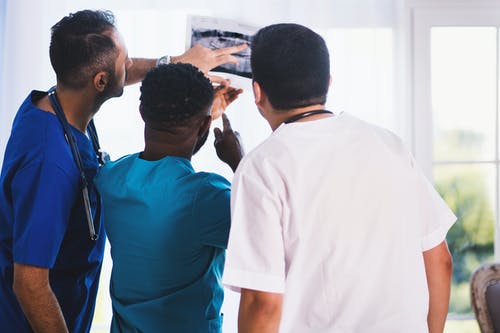
By definition, radiologists are medical professionals who utilize images, such as x-rays, to detect and diagnose illness. Their patients’ symptoms and possible diagnoses often guide doctors and primary care providers when deciding on the type of medical imaging studies to conduct.
Medical imaging technologists utilize their technical expertise and knowledge of human anatomy to capture precise images of specific areas of the body of patients, allowing doctors to check these parts for any signs of illness thoroughly. To ensure the best quality of patient care, radiologists evaluate these images and provide reports to referring clinical practitioners such as surgeons, pediatricians, obstetricians, and internists.
Diagnostic Imaging Methods
Radiology is one of the subfields of medicine crucial in various medical specialties like surgery, pediatrics and obstetrics, cancer treatment and trauma response, emergency medicine, infectious disease treatment, and many more. These are some of the more prevalent imaging diagnostic procedures utilized in medical procedures.
1. X-Ray Imaging
Imaging with X-rays is the most commonly used method for radiology imaging. A small dose of X-rays is administered to a specified body area to create an image called a radiograph. When used by radiologists and technologists trained properly, X-rays are generally secure.
X-rays are used to detect fractures. They also look at the various body parts like the chest, the abdomen, and the superficial soft tissues. They are an efficient and straightforward method for your doctor to identify different diseases in the body. For expert screening and diagnostic imaging in Austin, you can hit the web and search for facilities in your area.
2. Ultrasound
Sonography employs sound waves that are transmitted into the body. These are reflections by a transducer that produces an electrical signal that is then used to create a visual image. The method creates a live-feed video without radiation.
Doctors often use the use of ultrasound to track fetal growth and development. An ultrasound test could be an effective diagnostic tool if a patient is experiencing discomfort or swelling or suffering from an infection. Furthermore, medical professionals employ ultrasounds to look at the liver, kidneys, gallbladder, heart, and gallbladder.
3. Computed Tomography
CT scanning, also known as CAT scanning, is an imaging method for medical purposes that uses X-rays. It creates a series of cross-sectional images to study organs, tissues, and bones in greater detail.
Due to their more detail, computed tomography scans may be more effective than standard radiographs. However, the radiation they use is higher. CT scans can detect cancer, tumors, and internal injuries that result from an accident. Doctors can use CT scans to follow the patient’s healing from a broken limb or heart issue.
4. Magnetic Resonance Imaging
MRI employs superconducting magnetic and radio waves instead of radioactive ionization. If examining the tendon and ligaments, soft tissues, or organs, doctors usually opt to perform MRI. Brain MRI helps diagnose various ailments, such as cancer, stroke, eye conditions, and aneurysms.
Heart size, post-heart-attack damage, and vascular inflammation are just several cardiac ailments that MRI can evaluate. In addition, it detects liver, breast, ovary, kidney, pancreatic, and other malignancies. If searching for an MRI facility in Texas, you can type in “MRI in Austin, TX” for the best results.
5. Vascular Interventional Radiography
Vascular interventional radiology enables medical specialists to treat various ailments. They use angioplasty, stenting, thrombolysis, and other less invasive techniques. CT, ultrasounds, and X-ray fluoroscopy can be utilized in vascular interventional radiology.
Diseases of the blood vessels are treatable, problems with dilated or obstructed veins can be addressed, and benign tumor treatments can be targeted. The help vascular and neuro interventional radiology can help with the early diagnosis and treatment of strokes, aneurysms, and the like.